
A direct microscopic somatic cell count provides a precise method for detecting somatic cells in milk, which directly impacts dairy quality and mastitis detection rates. Recent studies confirm that increased somatic cell levels signal intramammary infections and trigger changes in milk composition, reducing yield and lowering cheese quality. Technicians must follow each step with care, as accuracy ensures reliable detection of subclinical mastitis before clinical symptoms appear. Many laboratories now use a somatic cell count tester to validate results, maintaining confidence in milk safety and farm profitability.
Key Takeaways
- Direct microscopic somatic cell count offers a cost-effective and accurate way to detect somatic cells in milk, helping identify mastitis early and maintain milk quality.
- Proper sample collection, mixing, storage, and slide preparation are crucial to ensure reliable and consistent somatic cell count results.
- Using modern somatic cell count tester alongside manual microscopy improves accuracy, speeds up testing, and reduces human error.
- Regular monitoring of somatic cell counts supports early mastitis detection, better milk quality, and compliance with international regulatory standards.
- Following standardized protocols and avoiding common errors through training and equipment calibration ensures trustworthy results for dairy quality control.
Overview of Direct Microscopic Somatic Cell Count
Method Summary
The direct microscopic somatic cell count remains a cornerstone in milk quality assessment and mastitis detection. This approach involves preparing a stained smear of milk on a microscope slide, then counting somatic cells under high magnification. Technicians can complete the process within an hour, making it suitable for rapid diagnostics in both laboratory and field settings. The method stands out for its cost-effectiveness, requiring only basic microscopy equipment and minimal consumables. It also allows for the observation of cell morphology, which helps differentiate between cell types and identify abnormal findings.
Despite these strengths, the direct microscopic somatic cell count method presents several limitations. Operators cannot distinguish between live and dead cells, which may lead to overestimation of cell counts. The process relies heavily on the skill of the technician, introducing subjectivity and potential for error. Uneven staining, cell clumping, and artifacts can further affect accuracy. Automated methods, such as fluorescent image cytometers or electronic counters, offer faster and more reproducible results, especially when handling large sample volumes. However, the direct microscopic approach remains valuable for detailed morphological analysis and in cost-sensitive environments. Many laboratories now use a somatic cell count tester alongside microscopy to validate results and improve reliability.
Tip: For best results, laboratories should combine the direct microscopic somatic cell count with automated validation using a somatic cell count tester, especially when processing high sample volumes or when regulatory compliance is required.
Required Equipment
Technicians must use precise and reliable equipment to ensure accurate somatic cell counts. The following table outlines the recommended specifications for microscopes and slides used in 2025:
| Specification Aspect | Recommended Features/Values |
|---|---|
| Microscope Type | Low magnification fluorescent microscope with automatic focusing |
| Imaging | CCD camera capturing stained cells |
| Image Capture | Up to 60 images per test with computer-controlled X:Y stage movements |
| Sample Volume (Slide) | Disposable measuring chambers (e.g., LACTOCHIP) with 8 μL sample volume |
| Cell Size Detection Range | 2 to 30 μm |
| Measurement Range | 10,000 to 10,000,000 cells/mL (max 20,000,000 cells/mL) |
| Slide Chamber Configurations | 2 or 4 chambers, horizontal or vertical orientation |
| Slide Chamber Depths | Variable depths ranging from 10 to 200 microns |
| Slide Grid Types | 10×10, Bürker, Fuchs Rosenthal, Neubauer Improved, Thoma grids |
| Operation Interface | 10-inch touchscreen tablet |
| Result Time | Rapid results within 20 to 60 seconds |
| Additional Features | Embedded printer, wireless keyboard and mouse, portable design |
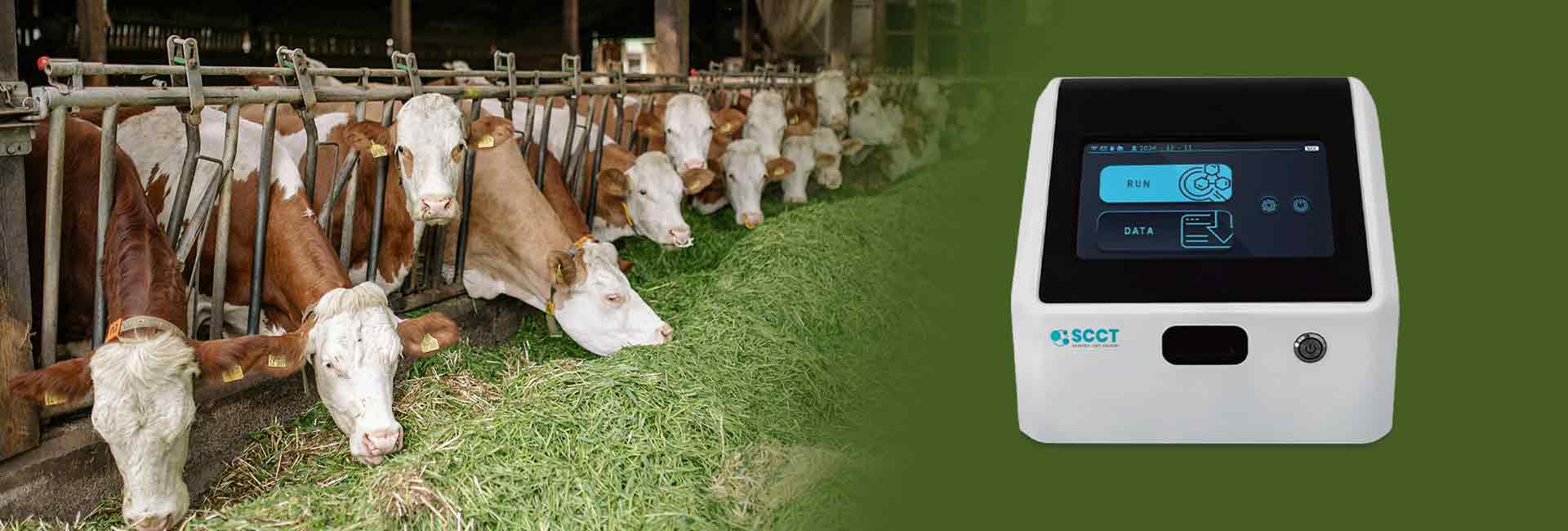
Modern somatic cell count testers, such as the LB-10SOC, detect cell sizes from 2 to 30 microns and capture up to 60 high-resolution images per test. These devices feature user-friendly touchscreens and deliver results in under a minute, making them ideal for real-time monitoring. Technicians can select from a variety of slide chamber configurations and grid types to match specific testing protocols. Disposable measuring chambers, like LACTOCHIP, help reduce contamination risk and streamline workflow.
Note: Proper equipment selection and maintenance play a critical role in achieving consistent and accurate direct microscopic somatic cell count results.
Sample Collection
Mixing and Handling
Accurate somatic cell count results begin with proper sample collection. Technicians should always prioritize cleanliness and consistency during this process. A well-mixed milk sample ensures that somatic cells distribute evenly, which is critical before preparing the smear. The industry standard recommends mixing the milk sample with 25 reciprocal movements. This action helps prevent cell clumping and provides a representative sample for analysis.
Best practices for collecting milk samples include the following steps:
- Wear clean, disposable gloves before handling any equipment or samples.
- Prepare each teat by wiping with a new alcohol wipe or alcohol-soaked cotton ball.
- Collect milk samples after teat preparation and before attaching the milking unit.
- Use sterile, unopened milk vials, avoiding contact between the vial and the teat end.
- Strip the teat until enough milk is collected for the sample.
- Label each sample clearly with cow identification, date, and quarter of collection.
- Immediately place samples on ice or refrigerate them.
- Follow any additional laboratory-specific instructions for storage and shipping.
Technicians should also discard the first streams of milk to reduce bacterial contamination. Consistent collection times and thorough labeling support reliable monitoring and traceability. Proper mixing and handling at this stage lay the foundation for accurate results when using a somatic cell count tester.
Storage and Transport
Proper storage and transport preserve the integrity of milk samples for somatic cell count analysis. Technicians should refrigerate samples at 4°C immediately after collection. This temperature maintains sample stability for up to two weeks, although testing within 72 hours remains ideal for optimal accuracy. Over extended periods, somatic cell counts may decrease, especially beyond the two-week mark. Freezing can extend sample life, but results may vary depending on milk type and laboratory protocols.
Samples should never be frozen unless specifically instructed by the laboratory. During transport, samples must remain chilled using ice packs or coolers. Technicians should avoid repeated temperature fluctuations, as these can compromise cell integrity. Prompt delivery to the laboratory ensures that the somatic cell count tester provides reliable and reproducible results.
Slide Preparation
Spreading the Sample
Accurate slide preparation begins with careful spreading of the milk sample. Technicians typically use 10 µL of milk, spreading it evenly over a 1 cm² area on a clean, degreased glass slide. This method ensures a representative distribution of somatic cells, which is essential for reliable microscopic analysis. Traditional glass slides sometimes cause uneven cell distribution due to edge effects, cell accumulation in the center, or air bubbles from coverslip placement. These issues can reduce observation accuracy and complicate counting.
Modern laboratories increasingly use microfluidic chips for this step. These chips employ controlled laminar flow, which eliminates edge effects and prevents bubbles. The nine-cell microfluidic chip design allows multiple observation cavities, enabling technicians to image separate areas and average the results. This approach improves accuracy and representativeness, especially when using a somatic cell count tester for validation.
Tip: Always prepare duplicate or quadruplicate slides to ensure repeatability and accuracy in somatic cell counts.
Drying and Fixing
Proper drying and fixing preserve the integrity of the milk smear and prevent cell loss during staining. The recommended procedure follows these steps:
- Spread 5–10 μL of fresh milk on a degreased slide, covering a 1 cm² area.
- Place the slide horizontally and allow the film to air dry.
- Fix the dried film by immersing it in 96% ethyl alcohol for 3 minutes.
- Air dry the slide again.
- Defat the slide with xylene for 10 minutes.
- Rinse with 60% ethyl alcohol and air dry.
- Keep the slide dustproof until staining.
Some protocols suggest cleaning the slide with ethanol, drying with lens paper, and flaming before use. Microinjection tools can help apply the milk sample evenly. Baking the slide on an electric stove may also be used to dry the film. These steps ensure that somatic cells remain fixed and visible for accurate counting, supporting reliable results when using a somatic cell count tester.
Consistent slide preparation techniques help maintain high standards in milk quality analysis and support regulatory compliance.
Staining
Stain Selection
Selecting the right stain ensures clear visualization of somatic cells under the microscope. In 2025, most laboratories prefer fluorescent stains or immunofluorescence reagents for their high sensitivity and specificity. These stains highlight cell nuclei and cytoplasm, making it easier to distinguish somatic cells from background debris. Technicians often choose stains compatible with automated imaging systems and somatic cell count tester validation. Popular options include acridine orange, DAPI, and specific antibody-based fluorescent dyes. Each stain offers unique advantages in terms of brightness, contrast, and compatibility with digital imaging.
Tip: Always verify stain compatibility with both the microscope and the somatic cell count tester to ensure accurate results.
Staining Steps
A standardized staining protocol improves reproducibility and accuracy in somatic cell counting. The following step-by-step procedure, adapted for 2025, supports both manual and automated workflows:
- Dewaxing (Day 1):
- Prepare twelve labeled staining boxes with xylene, xylene/ethanol, ethanol (100%, 95%, 70%), and distilled water.
- Incubate slides sequentially in xylene (three times, 15 minutes each), xylene/ethanol (5 minutes), 100% ethanol (twice, 2 minutes each), 95% ethanol (twice, 2 minutes each), 70% ethanol (twice, 2 minutes each), and distilled water (twice, 2 minutes each).
- Heat Antigen Retrieval:
- Place dewaxed slides in unmasking solution within a microwave-safe container.
- Microwave at 800 W until boiling, then continue boiling for 3 minutes.
- Cool slides in an ice box to approximately 50°C, then microwave again for 3 minutes.
- Allow slides to cool to room temperature.
- Initial Rinsing and Permeabilization:
- Mark slide edges with a hydrophobic barrier pen.
- Rinse slides in PBS twice for 2 minutes each, then for 10 minutes.
- Rinse in PBS/gelatin/Triton 0.25% twice for 10 minutes each.
- Blocking:
- Place slides in a humid box.
- Add 5% BSA solution and incubate for 60 minutes at room temperature.
- Primary Antibody Incubation:
- Remove blocking solution and add primary antibody.
- Incubate overnight in a humid box.
- Second Rinsing:
- Rinse slides in PBS twice for 10 minutes each, then in PBS/gelatin/Triton 0.25% for 10 minutes.
- Secondary Antibody Incubation:
- Add secondary antibody and incubate in a humid box.
- Final Rinsing and Mounting:
- Rinse slides in PBS three times for 10 minutes each, then in CuSO4/NH4Cl solution for 10 minutes.
- Rinse briefly with distilled water, dry slides, and add mounting medium.
- Cover with a coverslip, avoiding air bubbles, and dry horizontally in the dark for 24 hours.
This protocol ensures consistent staining, which supports accurate cell identification and counting. Laboratories using a somatic cell count tester benefit from this standardized approach, as it aligns with automated imaging requirements and regulatory standards.
Microscopic Examination
Oil-Immersion Use
Technicians achieve the highest accuracy in somatic cell identification by using oil-immersion microscopy. The recommended setup involves a fluorescence microscope with a ×10 eyepiece and a 60× oil immersion objective lens, such as the UPlanFI 60x/125 Oil. This configuration enhances resolution, allowing clear visualization of somatic cell nuclei in stained milk smears. Some laboratories also use a compound light microscope at 1000× magnification with oil immersion for this purpose. Immersion oil bridges the gap between the slide and the lens, reducing light refraction and sharpening cell details.
During examination, technicians focus on the stained nuclei, which appear as dark blue or fluorescent spots larger than eight microns. Cells smaller than four microns or those lacking a clear nucleus are excluded from the count. The ISO 13366-1 (2008) guidelines recommend counting only those cells that touch the top or bottom of the counting strip. This approach ensures consistency and prevents double-counting. When using a somatic cell count test kit for validation, technicians can compare manual counts with automated results to confirm accuracy.
Tip: Always apply immersion oil carefully to avoid air bubbles, which can distort the microscopic image and hinder accurate counting.
Counting Rules

Accurate somatic cell counts depend on strict adherence to established counting rules. Technicians must count only clearly identifiable somatic cells, excluding artifacts, debris, and any doubtful cells. Cells that appear clumped are counted as one unless two distinct nuclei are visible. Operator training remains essential, as misidentification can lead to significant errors.
The minimum number of cells to count varies with the concentration of somatic cells in the sample. The following table summarizes the recommended guidelines:
| Somatic Cell Concentration (thousands/mL) | Coefficient of Variation (CV%) | Minimum Number of Cells to Count |
|---|---|---|
| Less than 150 | 10 | 100 |
| 150 to 250 | 7 | 200 |
| 250 to 400 | 6 | 300 |
| 400 or more | 5 | 400 |
These rules follow statistical principles to ensure precision. Technicians should always reject doubtful cells to maintain accuracy. The somatic cell count tester can further support quality control by providing automated counts for comparison. Adhering to these protocols ensures reliable results for milk quality assessment and mastitis detection.
Calculation
Formula
Technicians calculate the somatic cell concentration in milk by applying a standardized formula. This formula ensures consistency across laboratories and supports regulatory compliance. The calculation for the direct microscopic somatic cell count uses the following equation:
Somatic Cell Count (cells/mL) = (Number of cells counted × Dilution factor × 1000) / Volume of milk spread (μL)
For example, if a technician counts 200 cells on a slide prepared with 10 μL of milk and no dilution, the calculation would be:
Somatic Cell Count = (200 × 1 × 1000) / 10 = 20,000 cells/mL
Technicians must ensure accurate measurement of the milk volume and consistent spreading on the slide. Any deviation can lead to significant errors in the final result. Laboratories often recommend counting multiple fields and averaging the results to improve precision.
Tip: Always verify the calibration of pipettes and use duplicate slides to minimize variability in the direct microscopic somatic cell count.
Using a Somatic Cell Count Tester
Modern laboratories increasingly rely on a somatic cell count tester to streamline the quantification process. These devices automate cell detection and counting, reducing human error and increasing throughput. The following table highlights the advantages of using a somatic cell count tester compared to traditional microscopic counting:
| Feature | Somatic Cell Count Tester | Traditional Microscopic Counting |
|---|---|---|
| Accuracy and Sensitivity | Advanced detection technologies, higher accuracy | Manual, lower sensitivity |
| Speed and Efficiency | Automated, rapid processing of many samples | Slow, manual, higher chance of error |
| Contamination Prevention | Disposable consumables, reduced cross-contamination | Higher risk due to manual handling |
| Application Scope | Dairy farms, factories, veterinary, research | Mainly laboratory settings |
Automated testers use technologies such as fluorescence flow cytometry and microfluidics, which deliver high sensitivity and specificity. Comparative studies show a concordance correlation coefficient above 0.95 between automated and manual methods, confirming the reliability of these instruments. The automation allows technicians to process more samples in less time, making the somatic cell count tester an essential tool for modern dairy operations.
Laboratories benefit from improved accuracy, efficiency, and contamination control when adopting automated somatic cell counting solutions.
Interpreting Results
Milk Quality
Somatic cell count (SCC) serves as a primary indicator of milk quality in dairy laboratories. Lower SCC values reflect healthy mammary tissue and minimal inflammation. According to international standards, milk with SCC below 100,000 cells/mL is considered normal and free from infection. Values above 200,000 cells/mL suggest possible inflammation or subclinical mastitis. Most countries set a practical upper limit for bulk milk SCC at 400,000 cells/mL, although some regions allow higher thresholds.
| SCC Threshold Category | SCC Value (cells/mL) | Description / Interpretation |
|---|---|---|
| Normal milk | < 100,000 | Considered normal milk with no infection |
| Indication of inflammation/infection | ≥ 200,000 | Threshold for subclinical mastitis (inflammation) |
| Practical tolerance upper limit | Up to 400,000 | Accepted upper limit for practical reasons |
| Country / Region | Legal Bulk Milk SCC Limit | Notes |
|---|---|---|
| European Union, Australia, New Zealand, Canada, Switzerland | 400,000 | Common international legal limit |
| South Africa | 500,000 | Slightly higher legal limit |
| USA | 750,000 | Higher legal limit |
| Brazil | 1,000,000 | Highest legal limit among listed countries |
Dairy processors and regulators use these thresholds to ensure product safety and quality. The method of direct microscopic somatic cell count provides reliable data for compliance. Many laboratories confirm results with a somatic cell count tester to maintain accuracy and meet export requirements.
Note: Consistently low SCC values support higher milk yields, better cheese production, and improved shelf life.
Mastitis Detection
Veterinarians and dairy managers rely on SCC to detect mastitis, especially subclinical cases that lack visible symptoms. Subclinical mastitis often appears when SCC exceeds 100,000 cells/mL. This threshold offers the highest sensitivity for early detection, reducing the risk of undiagnosed infections. Some protocols use 200,000 cells/mL for greater specificity, but this may miss early cases.
| SCC Threshold (cells/mL) | Sensitivity (Se) | Specificity (Sp) | Interpretation |
|---|---|---|---|
| 100,000 | Highest Se (~60.6%) | Lower Sp | Best for detecting subclinical intramammary infection (IMI) with fewer false negatives |
| 200,000 | Moderate Se | Higher Sp | Increased specificity but decreased sensitivity |
| 300,000 | Lowest Se (~20.3%) | Highest Sp | Most specific but least sensitive |
Subclinical mastitis detection depends on regular SCC monitoring. The direct microscopic somatic cell count method allows early identification of elevated SCC, prompting timely intervention. Clinical mastitis usually presents with even higher SCC, but visible symptoms guide diagnosis in those cases. Many dairy operations use a somatic cell count tester to automate detection and reduce human error.
Tip: Early detection of mastitis through SCC monitoring helps protect animal health and reduces economic losses.
Applications
Quality Control
Direct microscopic somatic cell count remains a cornerstone in dairy quality control programs. Laboratories use this method to measure white blood cells and epithelial cells in milk, providing a direct assessment of udder health and milk quality. Despite advances in automation, technicians still rely on this reference method to calibrate faster, instrumental systems. The process, often combined with methylene blue staining, serves as the international benchmark for evaluating milk from individual quarters, whole cows, or bulk tanks.
Routine quality control programs use direct microscopic somatic cell count to detect early signs of mastitis and monitor herd health. Technicians often validate results from automated systems, such as the somatic cell count tester, against this gold standard. This approach ensures accuracy and consistency across different testing platforms. While automated counters like Fossomatic or Coulter systems offer speed, they remain too complex and costly for on-farm use. The direct microscopic method, therefore, provides a practical and reliable solution for detailed analysis and ongoing calibration.
Laboratories that combine direct microscopic counts with somatic cell count tester validation achieve higher confidence in milk quality assessments.
Regulatory Use
Regulatory agencies worldwide set strict limits for somatic cell counts in milk to protect public health and ensure product quality. These standards vary by region, reflecting differences in market requirements and food safety priorities. The following table summarizes key regulatory limits in 2025:
| Region/Authority | Somatic Cell Count Limit (cells/mL) | Notes |
|---|---|---|
| European Union (EFSA) | 400,000 | Limit for raw milk |
| United States (FDA) | 750,000 | Legal limit; many processors use stricter standards |
| United States (Average) | ~300,000 | National average below federal threshold |
In 2025, the U.S. Agricultural Marketing Service introduced a draft certification program to align with European Union requirements for raw milk exports. This initiative aims to help U.S. producers meet the EU’s 400,000 cells/mL limit, which is stricter than the U.S. federal standard. Industry groups support these efforts but seek clarity on testing protocols and certification procedures. Regulatory agencies also promote comprehensive quality control systems, such as HACCP, to prevent unsafe milk from entering the food supply. The somatic cell count tester plays a vital role in meeting these regulatory demands by providing rapid, accurate results that support compliance and international trade.
Best Practices
Standardization
Standardization ensures that technicians achieve consistent and reliable somatic cell counts across different laboratories and testing environments. Leading dairy labs follow strict protocols for sample collection, slide preparation, staining, and counting. They use validated equipment, such as the somatic cell count tester, to minimize variability. Recent research highlights several key takeaways for best practices in somatic cell counting:
- The RT-10 meter provides the highest accuracy for individual quarter SCC at or below 200,000 cells/mL. It shows strong correlation with reference standards and delivers excellent sensitivity and specificity.
- The DSCC meter performs best for bulk tank milk SCC measurements at or below 100,000 cells/mL, offering reliable predictive accuracy.
- Validated meters help guide selective dry-cow therapy, which can reduce unnecessary antibiotic use and lower treatment costs.
- Common alternatives, including the California Mastitis Test, electrical conductivity meters, and infrared thermography, often lack sensitivity, specificity, or consistency. These methods are less reliable for quantitative SCC measurement.
- Experts recommend using quantitative, validated SCC measurement tools rather than qualitative or subjective tests to improve mastitis management and antibiotic stewardship.
Tip: Standard operating procedures and regular calibration of the somatic cell count tester help maintain accuracy and support regulatory compliance.
Avoiding Errors

Technicians can avoid common errors by following best practices at every stage of the process. Proper sample mixing, accurate pipetting, and careful slide preparation reduce the risk of uneven cell distribution. Using clean, degreased slides and high-quality stains prevents artifacts that could interfere with counting. Technicians should always count only clearly identifiable somatic cells and reject doubtful or clumped cells unless two nuclei are visible.
A checklist for error prevention includes:
- Verify calibration of all equipment, especially the somatic cell count tester.
- Prepare duplicate slides and average results for greater precision.
- Store and transport samples at recommended temperatures to preserve cell integrity.
- Train staff regularly to recognize and avoid common pitfalls.
Consistent attention to detail and adherence to standardized protocols ensure that somatic cell count results remain accurate and actionable for dairy quality control and mastitis detection.
Conclusion
Accurate somatic cell counting relies on careful sample handling, precise slide preparation, and strict adherence to standardized protocols. Regular testing supports early detection of intramammary infections and helps maintain milk quality, as studies show that routine monitoring and good hygiene practices improve udder health. New technologies, such as biosensors and the somatic cell count tester, offer rapid and reliable results. For those seeking advanced knowledge, international standards like ISO 13366-1 and resources from the IDF/ICAR project group provide valuable guidance.
FAQ
What Is the Main Purpose of a Somatic Cell Count In Milk?
A somatic cell count measures the number of white blood cells and epithelial cells in milk. This test helps detect mastitis and assess milk quality. Dairy labs often use a somatic cell count tester for fast and accurate results.
How Soon Should Milk Samples Be Tested After Collection?
Technicians should test milk samples within 72 hours of collection. Quick testing preserves cell integrity and ensures reliable results. Using a somatic cell count tester soon after sampling helps maintain accuracy.
Can a Somatic Cell Count Tester Replace Manual Microscopy?
A somatic cell count tester offers rapid, automated results and reduces human error. Many laboratories still use manual microscopy for confirmation, especially when cell morphology analysis is needed.
What Are Common Errors in Somatic Cell Counting?
Common errors include improper sample mixing, inaccurate pipetting, and misidentification of cells. Technicians should calibrate equipment, such as the somatic cell count tester, and follow standardized protocols to avoid mistakes.
Why Do Laboratories Use Duplicate Slides for Counting?
Duplicate slides improve accuracy and reliability. By averaging results from multiple slides, technicians reduce the impact of random errors. This practice supports quality control, especially when validating results with a somatic cell count tester.