
Direct microscopic somatic cell count in milk plays a critical role in evaluating milk quality. This method provides a direct assessment of somatic cells present in dairy samples. Direct microscopic somatic cell count offers insight into milk quality by helping detect inflammation such as mastitis. Dairy professionals rely on this approach to meet regulatory standards and maintain product quality. The ability to count cells directly allows for accurate monitoring of dairy health and supports the production of high-quality milk. Comparing direct microscopic somatic cell count in milk with other methods helps the dairy industry select the most effective tool for ensuring quality.
Key Takeaways
- Direct microscopic somatic cell count helps detect udder health issues like mastitis by counting cells directly under a microscope.
- This method is cost-effective and reliable, making it ideal for small dairy farms and labs with limited budgets.
- Skilled technicians can observe cell shapes to identify early signs of infection, improving milk quality and safety.
- Limitations include small sample size, difficulty distinguishing live from dead cells, and results depending on operator skill.
- Choosing the right testing method depends on accuracy needs, cost, speed, and available staff training.
Direct Microscopic Somatic Cell Count in Milk
Definition
Direct microscopic somatic cell count in milk refers to a laboratory technique that measures the number of somatic cell count present in a milk sample. Somatic cells include white blood cells and epithelial cells that originate from the udder tissue. Their presence in milk often signals udder health and can indicate mastitis, an inflammation that affects dairy cows. This method allows dairy professionals to assess milk quality directly by counting these cells under a microscope. Direct microscopic somatic cell count provides a reliable indicator for milk testing and helps ensure compliance with industry standards.
Method Overview
The process of direct microscopic somatic cell count in milk involves several precise steps. Each step ensures accurate results and supports effective milk testing for quality control.
- Collect milk samples and mix them thoroughly to prevent uneven cell distribution.
- Store and transport samples at recommended temperatures to preserve cell integrity.
- Prepare slides using clean, degreased glass slides to avoid artifacts.
- Apply high-quality stains to clearly differentiate somatic cells from other components.
- Pipette the milk sample accurately to maintain consistent volume on each slide.
- Prepare duplicate slides and average the counts for improved precision.
- Examine slides under a microscope, counting only clearly identifiable somatic cells. Exclude doubtful or clumped cells unless two nuclei are visible.
- Calibrate all equipment regularly, especially the somatic cell count tester, to maintain accuracy.
- Train technicians to recognize common errors and follow standardized protocols.
Note: Following these steps ensures that direct microscopic somatic cell count in milk provides reliable data for dairy quality control and mastitis detection.
Direct microscopic somatic cell count remains a cornerstone in milk testing. This method offers direct observation and quantification of somatic cells, which supports both routine quality checks and targeted mastitis investigations.
Advantages
Regulatory Acceptance
Direct microscopic somatic cell count stands as the official reference method for milk quality assessment in many regulatory frameworks. Authorities recognize this technique for its accuracy and reliability in determining somatic cell levels. Dairy laboratories use direct microscopic somatic cell count to validate results from automated instruments, ensuring compliance with safety and hygiene standards. This method supports regulatory monitoring by providing accurate somatic cell count score, which reflect udder health and milk quality. Reliable results from direct microscopic somatic cell count help dairy producers maintain high standards in milk production and management.
Regulatory bodies rely on direct microscopic somatic cell count for quality control and mastitis prevention. This method remains essential for dairy quality control and safety, especially when confirming low somatic cell count.
Cost-Effectiveness
Direct microscopic somatic cell count offers significant benefits in terms of cost-effectiveness. Unlike automated somatic cell count testers, which can cost between $2,200 and $4,200 per unit, direct microscopic methods require only basic laboratory equipment and skilled personnel. Many small dairy operations and laboratories choose this approach to reduce expenses while maintaining accuracy in milk quality assessment. Although automated devices like the ADAM-SCC provide affordable operation for some users, direct microscopic somatic cell count remains accessible for those with limited budgets.
Dairy producers benefit from lower initial investment and ongoing costs when using direct microscopic somatic cell count. This method allows regular monitoring of milk quality and udder health without the need for expensive machinery. The ability to achieve accurate results with minimal financial outlay supports sustainable milk production and management.
Cell Morphology Analysis
Direct microscopic somatic cell count enables detailed observation of cell morphology, which automated methods cannot provide. Technicians can distinguish between different types of somatic cells, such as white blood cells and epithelial cells, by examining their shape and structure under the microscope. This capability enhances the accuracy of milk quality assessment and supports early mastitis detection.
Studies show that direct microscopic somatic cell count serves as the gold standard for identifying subclinical mastitis and monitoring udder health. Automated methods may underestimate somatic cell counts, but microscopic analysis delivers reliable results by allowing direct visualization of cells. Dairy professionals use this method to detect changes in cell morphology that signal inflammation or infection, improving management and production outcomes.
Direct microscopic somatic cell count provides accurate somatic cell counts and supports early mastitis detection. The ability to observe cell morphology helps identify health issues before they affect milk yield and quality.
Key Benefits of Direct Microscopic Somatic Cell Count:
- Official reference method for regulatory compliance
- Lower cost compared to automated testers
- Accurate results for milk quality assessment
- Supports regular monitoring and dairy quality control
- Enables early mastitis detection and management
- Provides reliable results for udder health evaluation
- Allows observation of cell morphology for improved diagnosis
- Promotes hygiene and safety in milk production
Dairy producers and laboratories rely on direct microscopic somatic cell count to maintain high milk quality, ensure safety, and support effective management practices. This method delivers accurate results, supports regular monitoring, and helps prevent mastitis, leading to better milk yield and production quality.
Limitations
Sample Volume
Direct microscopic somatic cell count in milk uses a very small sample volume. This limitation can affect the accuracy of results, especially when the distribution of somatic cells is not uniform. A small sample may not represent the entire batch of milk, leading to possible errors in detecting low somatic cell count or high somatic cell count. When technicians analyze only a tiny portion, they might miss early signs of mastitis or subclinical mastitis. This challenge can impact udder health monitoring and mastitis prevention efforts.
Note: Small sample size increases the risk of missing abnormal cells, which can delay mastitis detection and affect milk quality.
Detection Limit
The detection limit of direct microscopic somatic cell count restricts its usefulness in some cases. This method struggles to identify very low somatic cell count in milk, which is important for confirming udder health and ensuring hygiene. When the cell count falls below a certain threshold, the results may not reflect the true condition of the milk. This limitation can hinder mastitis prevention and early intervention.
Direct microscopic methods also cannot distinguish between live and dead cells effectively. The technique focuses on counting total somatic cells, not assessing cell viability. Dead cells often lose their staining ability, making them harder to identify. Technicians rely on visual cues, but these can be subjective and inconsistent. Staining methods like trypan blue offer only a rough distinction and can introduce observer bias. High background signals or improper microscope settings may further interfere with accurate results. As a result, the method cannot reliably indicate whether the somatic cells in milk signal active infection or past inflammation.
- Challenges in distinguishing live and dead cells:
- Observer experience heavily influences accuracy.
- Morphological changes take time to appear.
- Staining methods provide only rough estimates.
- Misidentification can occur due to improper dye use.
- Background signals or debris can mask true results.
This limitation means that while the method can detect mastitis and subclinical mastitis, it cannot always provide a complete picture of udder health or the effectiveness of mastitis prevention strategies.
Operator Dependency
Operator dependency represents one of the most significant limitations of direct microscopic somatic cell count in milk. The accuracy and reproducibility of results depend on the skill and experience of the technician. Human error can introduce subjectivity, especially when identifying and counting somatic cells. Color artifacts, cell aggregation, and improper slide preparation can all affect results. Inconsistent technique may lead to variations in detecting low somatic cell count or high somatic cell count, which impacts udder health assessment and mastitis prevention.
The method is also time-consuming and requires skilled personnel. Technicians must prepare slides carefully, apply stains correctly, and count cells under a microscope. This process takes longer than automated methods and increases labor costs. Operator dependency makes it difficult to achieve consistent results across different laboratories or even within the same facility over time.
Operator dependency in direct microscopic somatic cell count can reduce accuracy and reproducibility. Automated or semi-automated methods, such as video microscopy, offer more consistent results and reduce the influence of human error.
Summary Table: Main Limitations of Direct Microscopic Somatic Cell Count
| Limitation | Impact on Results | Effect on Udder Health and Mastitis Detection |
|---|---|---|
| Small sample volume | May not represent whole batch | Misses early mastitis or subclinical mastitis |
| Detection limit | Struggles with low cell counts | Delays intervention and mastitis prevention |
| Operator dependency | Inconsistent, subjective results | Reduces reliability of health monitoring |
| Inability to distinguish live/dead cells | Incomplete health assessment | Limits mastitis diagnosis and prevention |
| Time-consuming process | Slower turnaround | Delays in hygiene and safety actions |
Direct microscopic somatic cell count in milk remains valuable for certain applications, but these limitations highlight the need for careful technique and skilled personnel. Dairy professionals must consider these factors when choosing a method for monitoring udder health, ensuring hygiene, and supporting mastitis prevention.

Comparison with Other Methods
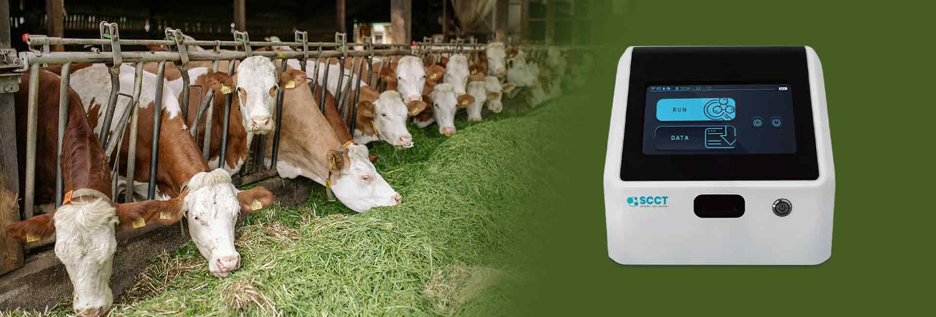
Somatic Cell Count Tester
Somatic cell count tester has become popular in many dairy laboratories. The device provides rapid and objective results for milk testing. The tester uses advanced technology to deliver accurate somatic cell counts, which helps producers monitor milk quality efficiently. Unlike DMSCC, somatic cell count test kit reduces operator dependency and minimize human error. Many testers can process large numbers of samples quickly, making them suitable for routine quality control.
However, somatic cell counter in milk may require regular calibration, especially when used for goat milk. Instruments calibrated for cow milk often overestimate somatic cell counts in goat milk unless adjusted for species differences. Non-infectious factors and natural variability in goat milk can affect accuracy. Despite these challenges, automated testers remain practical for high-throughput environments and deliver reliable results for most cow milk samples.
Flow Cytometry
Flow cytometry offers a modern approach to milk testing. This method uses DNA-specific fluorescent dyes to identify and count somatic cells. Flow cytometry provides rapid, objective, and highly accurate results. It can differentiate between viable and non-viable cells, which improves the assessment of udder health. Laboratories value this method for its ability to handle complex milk samples, especially those with high levels of cytoplasmic particles, such as goat milk.
Flow cytometry stands out for its speed and repeatability. It avoids the subjectivity of DMSCC and delivers reliable results even when cell numbers are low. The method requires specialized equipment and reagents, but it supports routine testing and quality monitoring. Flow cytometry also enables quick determination of differential cell counts, which enhances the accuracy of mastitis detection.
Flow cytometry is especially useful for goat milk, where cytoplasmic particles can interfere with other methods. DNA-specific staining ensures accurate somatic cell counts and reduces misidentification.
Chemical Methods
Chemical methods, such as the California Mastitis Test, provide a simple and cost-effective way to screen milk for high somatic cell counts. These tests use reagents that react with cell components, producing visible changes. Chemical methods offer quick results and require minimal equipment, making them suitable for on-farm testing.
While chemical methods are easy to use, they lack the accuracy and sensitivity of DMSCC, automated testers, or flow cytometry. They provide only an estimate of cell concentration and cannot distinguish cell types or viability. Chemical tests serve as useful screening tools but do not replace laboratory-based methods for precise milk quality assessment.
Comparison Table: Key Features of Somatic Cell Count Methods
| Method | Speed | Accuracy | Practicality | Suitable For | Reliable Results |
|---|---|---|---|---|---|
| DMSCC | Slow | High* | Laborious | Small labs, research | Yes* |
| Automated Tester | Fast | High | Practical | Routine testing | Yes |
| Flow Cytometry | Fast | Very High | Advanced | Complex samples | Yes |
| Chemical Methods | Very Fast | Low | Simple | On-farm screening | No |
*DMSCC provides accurate results when performed by skilled personnel but may lack repeatability.
Choosing a Somatic Cell Count Tester
Selection Factors
Selecting the right somatic cell count tester is important for maintaining milk quality and supporting dairy production. Several factors influence this decision, each affecting regular monitoring, management, and milk yield.
Key factors to consider include:
- Accuracy
Laboratories and dairy producers need reliable results for milk quality and udder health. Direct microscopic methods offer high accuracy when technicians count enough cells. Automated testers provide fast and reproducible results, but some may have limitations with certain milk types or particle sizes. - Cost
Budget plays a major role in choosing a method. Direct microscopic counting requires a basic microscope and costs less than automated or flow cytometry devices. Automated counters and flow cytometers can be expensive, but they save time and labor in large-scale dairy production. - Practicality
The practicality of a method depends on the size of the dairy operation and the need for regular monitoring. Small farms may prefer affordable, manual methods. Large dairies often choose automated testers for faster milk testing and higher throughput. - Speed and Throughput
Automated methods process many samples quickly, supporting efficient milk production and regular monitoring. Manual counting takes more time and may slow down dairy quality control. - Labor and Training
Direct microscopic counting needs skilled personnel and careful slide preparation. Automated and flow cytometry methods reduce operator dependency, making them suitable for routine milk quality checks in busy labs.
Experts recommend that laboratories select a method based on available resources, desired accuracy, and the context of milk production. Increasing the number of cells counted in direct microscopic methods improves accuracy and reduces result variation. Automated testers offer rapid, reproducible results, making them ideal for high-volume dairy management.
Comparison of Somatic Cell Count Methods
| Method | Advantages | Limitations | Expert Recommendations |
|---|---|---|---|
| Direct Microscopic Counting | Affordable, reliable, accurate with enough cells | Time-consuming, human error, needs skilled staff | Good for routine use if cell count per assay is increased |
| Somatic Cell Count Tester | Fast, efficient, moderate precision | Expensive, calibration needed, particle size limits | Suitable for rapid, reproducible results in large-scale production |
| Flow Cytometry | Highly reproducible, precise | Costly, technical limits, bulky equipment | Best for reproducibility, less accurate for some milk samples |
Dairy professionals should match their choice to the needs of their operation. Regular monitoring, milk yield, and overall management benefit from a method that balances accuracy, cost, and practicality. The right tester supports high milk quality and efficient milk production.

Conclusion
Direct microscopic somatic cell count in milk offers clear benefits for milk quality assessment, udder health evaluation, and mastitis detection. This method provides accurate results and supports regulatory compliance, as mandated by federal standards for both cow and goat milk. Dairy producers use DMSCC to confirm milk quality and regain compliance when somatic cell counts fall below set thresholds. While DMSCC requires skilled personnel and careful technique, it remains valuable for small dairy operations and situations needing detailed cell analysis. Automated methods suit high-volume milk testing, but DMSCC excels in precise health monitoring and early mastitis detection.
FAQ
What Is the Main Purpose of Direct Microscopic Somatic Cell Count In Milk?
Direct microscopic somatic cell count in milk helps dairy professionals assess milk quality and udder health. This method supports early mastitis detection and provides accurate somatic cell counts. Reliable results guide dairy management and ensure compliance with hygiene and safety standards.
How Does Direct Microscopic Somatic Cell Count Benefit Dairy Quality Control?
This method allows regular monitoring of milk for high somatic cell count or low somatic cell count. Accurate results help identify subclinical mastitis and support mastitis prevention. Dairy quality control improves when producers use this approach for milk quality assessment and management.
Why Is Operator Skill Important for Accurate Results?
Operator skill affects the accuracy and reliability of direct microscopic somatic cell count. Skilled technicians produce consistent results and minimize errors. Proper training ensures that dairy laboratories maintain high standards for milk testing, milk production, and udder health monitoring.
Can Direct Microscopic Somatic Cell Count Detect Early Mastitis?
Yes. Direct microscopic somatic cell count enables early mastitis detection by identifying increases in somatic cells. Early intervention helps protect milk yield, maintain milk quality, and support mastitis prevention. Regular monitoring with this method benefits overall dairy health and production.
What Are the Limitations of This Method Compared To Automated Testing?
Direct microscopic somatic cell count requires more time and skilled personnel. Small sample volume and operator dependency can affect results. Automated testing offers faster, more consistent results for large-scale milk production, but may lack detailed cell morphology analysis.