
Teat-end hyperkeratosis increases risk of SCC in older cows by weakening the udder’s natural defense. Thickened tissue at the teat end creates openings for bacteria to enter, which raises the chance of mastitis. Severe callosity at the teat end often prevents proper closure of the teat canal and further heightens the risk of clinical mastitis. Machine milking can worsen these changes in teat condition. Regular use of a somatic cell count tester helps identify problems early and supports better udder health.
Key Takeaways
- Teat-end hyperkeratosis weakens the udder’s defenses, increasing the risk of mastitis and higher somatic cell counts.
- Regular monitoring with a somatic cell count tester helps identify issues early, allowing for prompt intervention and better udder health.
- Proper milking techniques and machine settings are crucial to prevent teat-end damage and reduce the risk of hyperkeratosis.
- Maintaining good hygiene and providing clean bedding significantly lowers the risk of infections and supports milk quality.
- Dairy producers should assess at least 20% of the herd’s teat ends regularly to monitor health and prevent mastitis.
Teat-End Hyperkeratosis and SCC Risk
What Is Teat-End Hyperkeratosis?
Teat-end hyperkeratosis describes the thickening and hardening of the skin at the end of the teat in dairy cows. This condition often appears as raised, smooth, or rough rings around the teat ends. One or more teats can show these changes in a single cow. Dairy herds frequently observe this issue, especially in cows exposed to repeated machine milking or environmental stress. The development of hyperkeratosis signals a breakdown in the natural barrier that protects the udder from infection.
- Raised, rough, or smooth rings at the teat end
- Multiple teats may be affected in one cow
Teat-end hyperkeratosis stands out as a significant risk factor for udder health problems in dairy cows. The thickened tissue can prevent the teat canal from closing properly, which increases the risk of bacterial invasion and subsequent mastitis.
How Hyperkeratosis Affects Somatic Cell Count?
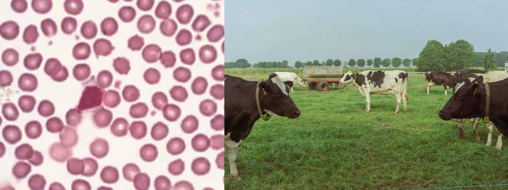
Hyperkeratosis impacts the somatic cell count in milk by altering the teat’s natural defense. When the teat end becomes callused, bacteria can enter the udder more easily. This triggers an immune response, causing the somatic cell count to rise. Research shows a direct relationship between the severity of hyperkeratosis and elevated SCC in dairy cows.
| Study | Findings |
|---|---|
| Gleeson et al. (2004) | Investigated the effect of hyperkeratosis on somatic cell counts in dairy cows, indicating a direct relationship. |
| Neijenhuis et al. (2001) | Explored the relationship between teat-end callosity and clinical mastitis occurrence, which correlates with elevated somatic cell counts. |
Management practices also play a role. Improved hygiene and pre-dipping can lower SCC, even in cows with hyperkeratosis. However, severe cases of hyperkeratosis remain a risk for increased SCC and mastitis.
| Finding | Description |
|---|---|
| Teat-end hyperkeratosis | Not a risk factor for high SCC in all cases |
| Management practices | Improved hygiene and pre-dipping can lower SCC |
Environmental factors, such as high pathogen loads and poor weather, can worsen hyperkeratosis and further increase SCC. The use of inappropriate disinfectants or emollients may also damage the teat end, raising the risk of infection and higher somatic cell counts.
Link to Clinical Mastitis
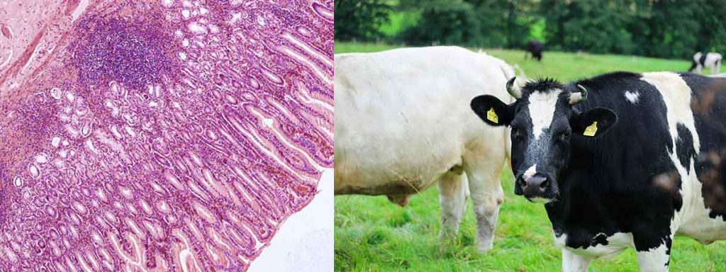
Teat-end hyperkeratosis directly links to an increased risk of clinical mastitis in dairy cows. The thickened, rough surface at the teat end allows bacteria to colonize and enter the udder. Studies show that cows with severe hyperkeratosis have a much higher risk of developing clinical mastitis, especially from pathogens like E. coli and Strep. uberis.
| Evidence Description | Findings |
|---|---|
| Higher incidence of clinical mastitis in cows with hyperkeratinized teat ends | Related to high contamination of the teat ends by bacteria and infiltration into glandular tissue. |
| Increased microbial load in teats with callused ends compared to normal teats | Indicates that hyperkeratosis leads to a higher risk of bacterial entry and subsequent mastitis. |
| Continuous destruction and regeneration of cells in hyperkeratosis | Results in thickening of tissue, negatively influencing teat canal closure and increasing microbial load. |
| Study Focus | Findings |
|---|---|
| Risk factors associated with clinical mastitis | Increasing severity of teat-end hyperkeratosis correlates with a higher risk of clinical mastitis, particularly for E. coli and Strep. uberis. |
| Relationship between teat-end callosity and mastitis | Cows with clinical mastitis had more teat-end callosity than healthy cows, especially during specific lactation months. |
| Impact of hyperkeratosis severity | Cows with very severe hyperkeratosis had a higher risk of clinical E. coli and Strep. uberis mastitis. |
| Risk factors for clinical mastitis | Quarters with very severe hyperkeratosis were significantly more likely to develop clinical mastitis compared to those with moderate or thin callosity. |
Severe teat-end hyperkeratosis serves as a risk factor for both clinical and subclinical mastitis. Once the protective layer at the teat end is compromised, pathogens such as Staphylococcus aureus can colonize the tissue, leading to infection. Environmental hygiene, weather conditions, and management practices all influence the risk of hyperkeratosis and subsequent mastitis in dairy cows.
Dairy producers must recognize the importance of monitoring teat condition and somatic cell count to reduce the risk of mastitis and maintain udder health. Regular assessment of teat ends and prompt action can help lower the incidence of clinical mastitis and improve milk quality in dairy herds.
Older Cows and Mastitis Risk
Age-Related Teat Changes
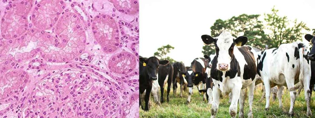
Older cows in dairy herds experience significant changes in teat structure as they age. The teat becomes longer and wider, which increases the risk of hyperkeratosis. These changes make the teat end more vulnerable to damage and infection. As the udder ages, the tissue loses elasticity, and the teat canal may not close as tightly. This breakdown in the natural barrier allows bacteria to enter the udder more easily, raising the risk of mastitis. Increased lactation number also contributes to the development of teat-end callosity.
| Evidence Type | Findings |
|---|---|
| Study Population | 4,022 German Holstein cows were studied. |
| Associations | Teat-end hyperkeratosis is associated with lactation number, teat length, and teat diameter. |
| Recommendations | Further research is needed to reduce the prevalence of teat-end hyperkeratosis in high-yielding dairy herds. |
Prolonged Milking Effects
Prolonged milking sessions in dairy cows lead to changes in teat-end tissue. Longer suction phases from milking machines increase the risk of very severe hyperkeratosis. Machine settings and techniques play a major role in the development of teat-end callosity. Improper milking techniques can damage the teat ends, making the udder more susceptible to mastitis.
| Evidence Type | Findings |
|---|---|
| Tissue Changes | Prolonged milking duration leads to changes in teat-end tissue, resulting in hyperkeratosis. |
| Machine Techniques | Milking machine techniques influence both short-term and long-term teat conditions. |
| Suction Phases | Longer suction phases can increase teat-end callosity. |
| Factors | Prevalence of teat-end hyperkeratosis is associated with lactation number, teat size, and machine settings. |
Tip: Regularly checking machine settings and adjusting milking duration can help reduce the risk of very severe hyperkeratosis and protect udder health.
Increased Susceptibility to Clinical Mastitis
Older cows face a higher risk of clinical mastitis compared to younger cows. Increased parity means more lactation cycles, which weakens the udder’s defenses. Physiological changes in aging cows lower their resistance to intramammary infections. Management practices often favor younger cows, leaving older cows at greater risk. Improper milking techniques further increase the risk of quarter case of clinical mastitis, especially in cows with very severe hyperkeratosis.
| Evidence Type | Description |
|---|---|
| Parity Impact | Older cows show a higher incidence of clinical mastitis due to increased parity, indicating that as cows age, their risk of infection rises. |
| Management Practices | Younger cows are often managed with higher standards, reducing their risk of clinical mastitis compared to older cows. |
| Physiological Changes | Aging cows may experience a decline in resistance to intramammary infections (IMI), making them more susceptible to clinical mastitis. |
- Improper milking techniques can worsen teat health and hygiene, increasing the risk of infections.
- Severe teat-end hyperkeratosis is linked to both clinical and subclinical mastitis due to higher susceptibility to pathogens.
- Damaging the teat ends through improper milking can lead to increased infection likelihood.
Older dairy cows with very severe hyperkeratosis and repeated exposure to improper milking face the highest risk of mastitis. Monitoring teat-end callosity and udder condition in these cows helps reduce the risk of quarter case of clinical mastitis and supports better milk quality.
Managing Somatic Cell Count and Mastitis
Monitoring SCC with Somatic Cell Count Tester
Dairy producers rely on regular monitoring to control mastitis and maintain milk quality. A somatic cell count tester provides fast and accurate results, helping identify cows with elevated cell counts before clinical signs appear. Monitoring bulk tank somatic cell count gives an overview of herd health. Producers often separate infected cows to prevent the spread of infection. Clean bedding and consistent pre- and post-milking routines reduce environmental infection risk. Reviewing DHIA reports for linear cell scores and infection percentages helps track trends over time. Culturing milk samples identifies specific pathogens, allowing for targeted management strategies. Proper teat dip coverage and cleanliness during milking lower the risk of bacterial infection and keep cell counts in check.
Tip: Early detection of mastitis through regular use of a somatic cell count tester allows for prompt intervention and reduces the risk of severe infection.
Preventing Hyperkeratosis and Mastitis
Prevention remains the most effective strategy for reducing mastitis and improving udder health in dairy cows. Teat dipping with softening agents after each milking session supports healing and protects the teat end. Effective teat dipping at the end of milking lowers the risk of new intramammary infections. Proper milking machine settings, including correct vacuum and pulsation, prevent excessive pressure on the teat and reduce hyperkeratosis. Shorter milking times with optimal flow rates also help minimize tissue damage. Environmental management, such as reducing cow stress and maintaining hygiene, further decreases the risk of infection.
| Factor | Description |
|---|---|
| Milking Machine Settings | Increased pressure from teat cup liners contributes significantly to hyperkeratosis. |
| Milking Time | Longer milking times with low flow rates increase hyperkeratosis incidence. |
Dairy herds benefit from internal teat sealants, which protect the udder until natural keratin seals the canal. Good hygiene scores after the dry period lower infection risk. Genetic selection for lower somatic cell score and increased mastitis resistance improves herd health over time.
- Teat end scoring helps assess teat health and the effectiveness of milking procedures.
- Scoring should include at least 80 cows or 20% of the herd, performed quarterly and after changes in milking routines.
- Veterinarians play a key role in interpreting results and guiding management decisions.
Improving Milk Quality and Udder Health
Improving milk quality and udder health requires a combination of good hygiene, proper milking technique, and regular monitoring. More than 20% of cows with poor udder hygiene scores face increased mastitis risk and lower milk quality. Bedding maintenance and overall farm hygiene have a strong influence on somatic cell count in milk. Lack of teat disinfection after milking leads to higher cell counts and more cases of subclinical mastitis, which negatively affects milk quality.
| Benefit | Description |
|---|---|
| Improved Milk Quality | Low cell counts result in better milk products, longer shelf life, and improved flavor. |
| Increased Milk Yield | Cows with low somatic cell counts produce more milk, boosting dairy productivity. |
| Reduced Treatment Costs | Fewer infections mean lower veterinary expenses for the dairy operation. |
| Enhanced Herd Health | Regular assessment of cell counts helps manage udder health and reduce milk losses. |
| Better Consumer Confidence | Maintaining low cell counts builds trust and supports sales in the global market. |
| Improved Cow Welfare | Good management of cell counts contributes to cow comfort and overall welfare. |
Note: Early detection of mastitis in cows with a history of teat-end hyperkeratosis is critical. Severe keratin frond growth at the teat end increases the risk of clinical mastitis by more than seven times compared to cows with normal teat ends. Barn hygiene and previous mastitis history also serve as important indicators for early intervention.
Dairy workers benefit from training programs that teach proper teat end scoring, assessment of hardness, color changes, swelling, and abrasions. Following guidelines from organizations like Teat Club International ensures consistent monitoring and better management of mastitis risk.
Maintaining low somatic cell counts supports high milk quality, increased yield, and better cow welfare. Proactive management, regular use of a somatic cell count tester, and attention to hygiene and milking practices protect both the udder and the dairy’s bottom line.
Conclusion
Teat-end hyperkeratosis increases SCC and mastitis risk in older cows by weakening the udder’s natural defenses. Dairy herds can protect milk quality and udder health through proactive management. Experts recommend these steps:
- Check weather forecasts and use a weather station for quick decisions.
- Evaluate at least 20% of the herd’s teat ends after milking.
- Maintain a strong mastitis prevention program with proper sanitation.
- Provide clean, dry bedding and adequate housing.
Regular monitoring with a somatic cell count tester supports early detection and better outcomes.
FAQ
What Causes Teat-End Hyperkeratosis in Dairy Cows?
Teat-end hyperkeratosis develops from repeated machine milking, improper milking techniques, or environmental stress. Older cows show more changes due to age and repeated lactations. Poor hygiene and harsh weather also contribute to the condition.
How Does Hyperkeratosis Affect Milk Quality?
Hyperkeratosis increases the risk of bacterial entry into the udder. This leads to higher somatic cell counts and more cases of mastitis. High cell counts lower milk quality and reduce shelf life.
Can Farmers Prevent Teat-End Hyperkeratosis?
Farmers can prevent hyperkeratosis by adjusting milking machine settings, using proper milking routines, and maintaining good hygiene. Regular teat scoring and early intervention help protect teat health.
Why Do Older Cows Have Higher SCC Levels?
Older cows have longer and wider teats. Their teat canals do not close as tightly. These changes make them more prone to infection and higher somatic cell counts.
What Tools Help Monitor Udder Health?
A somatic cell count tester gives fast results for detecting udder problems. Farmers also use teat scoring, milk culturing, and DHIA reports to track trends and manage herd health.