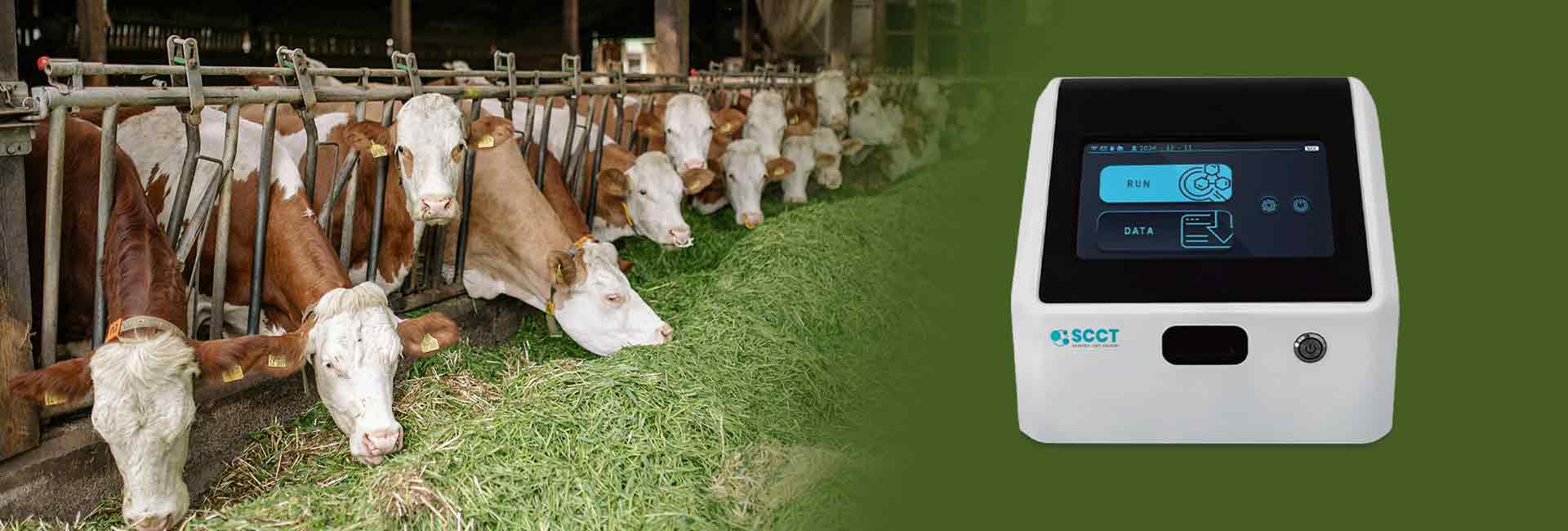
Accuracy in cell counting depends on advanced technology and strict operational protocols. The somatic cell counter uses digital imaging and flow cytometry to address common sources of error, such as manual microscopy and indirect methods.
Advancement Type
Contribution to Accuracy
Integration of AI and machine learning
Enhances accuracy and reduces manual errors
Implementation of digital imaging
Improves precision and enables detailed cellular analysis
Adoption of flow cytometry-based counters
Allows for multiparametric analysis
Key Takeaways
Advanced technology, like AI and flow cytometry, enhances accuracy in somatic cell counting by reducing manual errors.
Proper sample preparation, including cleaning and mixing, is crucial for reliable results and prevents inaccuracies caused by clumping.
Optimizing device settings, such as chamber height and counting parameters, ensures precise measurements tailored to each sample type.
Regular calibration and maintenance of cell counting instruments are essential for consistent performance and accurate results.
Choosing the right cell counting method impacts the reliability of results, with automated methods generally providing greater accuracy than manual techniques.
Sample Preparation
Clean Surface
A clean sample surface plays a vital role in accurate somatic cell counting. Technicians must remove any debris or contaminants before analysis. This step prevents interference with imaging and ensures that the sample reflects true cell numbers. Washing teats with water before milking enhances overall milk quality and improves the sample. Regular cleaning of milking lines also contributes to better sample quality. Strip cup tests help assess somatic cell levels and identify potential issues. The California mastitis test can detect problems before milking begins. These practices support the goal of obtaining a clean sample surface for reliable results.
Mixing Techniques
Proper mixing techniques help distribute cells evenly throughout the sample. Technicians should mix the sample immediately before loading it into the somatic cell counter. This action helps minimize cell clumping and ensures that each portion contains a representative number of cells. Uneven mixing can cause inaccurate counts and reduce reliability. Consistent agitation of the sample maintains homogeneity and supports precise analysis. Mixing also prevents cells from settling at the bottom, which could skew results.
Clumping Prevention
Clumping prevention is essential for accurate cell counting. When cells clump together, the counter may underestimate the true cell concentration. Immediate mixing before loading the sample helps minimize cell clumping. Technicians must use consistent sample volume and proper loading techniques to achieve reliable results. The following practices improve sample handling:
Regular cleaning of milking lines
Conducting strip cup tests
Performing the California mastitis test
Washing teats with water before milking
These steps ensure that the sample remains uniform and free from clumps. Reliable sample preparation leads to trustworthy somatic cell counts.
Somatic Cell Counter Settings
Chamber Height
Chamber height selection plays a crucial role in the performance of a somatic cell counter. The chamber height determines the volume of the sample that the device analyzes during each run. A correct chamber height ensures that the cell counting method produces results with high precision. If the chamber is too shallow, the device may not capture enough cells, leading to underestimation. If the chamber is too deep, overlapping cells can cause inaccurate counts. Technicians must select the chamber height based on the expected cell concentration and the type of sample. The somatic cell count tester offers adjustable chamber heights to match different sample requirements. This flexibility supports accurate and repeatable measurements.
Tip: Always verify the chamber height setting before starting a new analysis. This simple step helps maintain consistency and reduces the risk of error.
Parameter Adjustment
Adjusting parameters is essential for optimizing the cell counting method. Each sample type may require different settings to achieve the best results. The somatic cell counter allows users to adjust counting parameters such as diameter, roundness, and threshold. These parameters help the device distinguish between somatic cells and other particles. For example, the recommended diameter range for CHO cells is 4–24 µm when using AO/PI staining and 8–30 µm with Trypan Blue. Roundness and threshold values also vary depending on the staining method and cell viability.
Parameter
Description
Recommended Range for CHO Cells
Diameter
Total size range of relevant cells expressed as min and max values.
4—24 µm (AO/PI), 8—30 µm (Trypan Blue)
Roundness
Determines relevant cell morphology for different channels.
1 (AO/PI), 60 (live), 25 (dead) (Trypan Blue)
Threshold
Level of intensity required for inclusion in a count.
1 (green/red for AO/PI), 35 (Trypan Blue)
Sampling frequency also impacts the accuracy of the cell counting method. Dynamic sampling every 24 hours, as shown in studies by Sorensen et al., improves test performance compared to fixed frequencies. The somatic cell count tester uses automated counting of stained cell nuclei, which correlates strongly with laboratory SCC measures (R² up to 0.99). This high correlation supports the use of automated counters for mastitis detection and routine monitoring.
Optimized Settings
Optimizing device settings for each analysis ensures the highest level of precision and efficiency. The somatic cell counter uses advanced image-processing algorithms and computer vision models to transition from qualitative to quantitative counting. Automation of image analysis eliminates manual assessment, which increases repeatability and throughput. The integration of a web-based user interface makes the somatic cell count tester accessible to users without specialized training.
Evidence Description
Impact on Accuracy and Throughput
Use of image-processing algorithm in AVM reader
Enhances accuracy and efficiency by enabling quantitative counting.
Automation of image analysis
Ensures greater repeatability and efficiency.
Integration of computer vision model
Significantly improves precision of SCC measurements.
Web-based UI development
Makes the device accessible for non-specialized users.
Validation against flow cytometry
Confirms laboratory-grade accuracy with strong correlation (>90%).
Field trials results
Demonstrates high sensitivity and specificity in real-time analysis.
Field trials have demonstrated that optimized settings lead to high sensitivity and specificity, making the somatic cell counter suitable for real-time analysis in various environments. Validation against flow cytometry confirms that the device achieves laboratory-grade accuracy with minimal bias. These features ensure that the cell counting method remains reliable and precise, regardless of the sample type or testing environment.
Note: Regular review and adjustment of device settings help maintain optimal performance and support consistent results.
Cell Counting Methods
Brightfield Imaging
Brightfield imaging remains a traditional method for somatic cell counting. This technique uses standard light microscopy to visualize cells in a sample. Many laboratories rely on brightfield imaging because it is straightforward and cost-effective. However, this method can introduce subjectivity, as technicians must manually identify and count cells. Manual counting often leads to variability in results, especially when different users analyze the same sample. Automated cell counters improve repeatability and reduce user error, but brightfield imaging still lacks the sensitivity of more advanced techniques.
Fluorescence Imaging
Fluorescence imaging uses fluorescent dyes to label somatic cells, making them easier to detect and count. This method increases sensitivity and allows for the identification of specific cell types. Automated counters equipped with fluorescence imaging provide more consistent results compared to manual methods. The use of fluorescent stains helps distinguish somatic cells from debris, which improves accuracy. Laboratories often choose fluorescence imaging when they require precise results, especially for samples with low cell concentrations. The evolution of somatic cell counting now includes smartphone-based testing, which uses fluorescence imaging for rapid and portable results. This approach offers cost-effectiveness and efficiency, making it suitable for dairy farms and small laboratories.
Flow Cytometry
Flow cytometry stands out as a widely adopted automated method for somatic cell counting. This technology analyzes thousands of cells per second, providing high-throughput results. Flow cytometry uses lasers and detectors to measure cell properties, offering rapid characterization and absolute cell counts. However, some limitations exist, such as challenges in measuring precise intake volumes and potential calibration issues. The method does not provide spatial information about cells. The table below summarizes the advantages and limitations of flow cytometry:
Advantages
Limitations
High throughput analysis
Challenges in measuring precise intake volumes
Rapid characterization of cells
Potential inaccuracies due to calibration
Absolute cell counts
Lack of spatial information
The choice of cell counting method affects the accuracy and reliability of results. Calibration issues can cause automated counters to overestimate somatic cell counts in certain species. Reliability varies between equipment unless properly calibrated. Accuracy and reliability decrease when results exceed 1.5×10^6/ml. Laboratories consider factors such as cost, throughput, efficiency, and repeatability when selecting a method. Automated counters offer high throughput and standardized results, while manual methods require more training and can introduce user error.
Note: Selecting the right cell counting method ensures reliable results and supports effective monitoring of milk quality and udder health.
Accuracy Validation
Calibration
Calibration stands as a critical step for maintaining the accuracy of cell counting instruments. Technicians calibrate the somatic cell count tester at regular intervals to ensure that measurements remain consistent and reliable. Calibration involves using reference samples with known cell concentrations. The instrument compares its readings to these standards and adjusts its algorithms to minimize error. This process helps validate the precision of the instrument and supports reproducible results.
A well-calibrated instrument produces accurate results across different sample types. Calibration also reduces the risk of bias caused by environmental factors or instrument drift. Technicians document calibration data to track instrument performance over time. The use of sophisticated software algorithms allows the somatic cell count tester to self-correct and maintain high standards of accuracy.
Regular calibration ensures that the instrument delivers reproducible results and supports laboratory-grade quality.
Maintenance
Routine maintenance plays a vital role in preserving the performance of cell counting instruments. Technicians clean and inspect the instrument to prevent clumping and contamination. Maintenance tasks include checking fluidics, replacing worn components, and updating software algorithms. These actions help validate the performance of the somatic cell count tester and minimize variability in data.
Common challenges in maintaining instrument accuracy include environmental pathogens, housing conditions, and hygiene standards. The table below summarizes these challenges and their solutions:
Employ trained staff and maintain high standards of hygiene and animal husbandry.
Impact of housing conditions on udder health
Frequent renewal of straw and complete cleaning of housing areas.
Necessity for high standards of hygiene
Use both objective and subjective scoring methods to monitor cow hygiene and somatic cell counts.
Cow cleanliness in organic dairy herds significantly affects somatic cell counts. A scoring system for cow hygiene can predict reductions in somatic cell counts. Maintenance routines also include monitoring for clumping, which can interfere with measurements and reduce reproducible results. Technicians use both objective and subjective scoring methods to assess instrument and sample quality.
Tip: Scheduled maintenance and hygiene protocols help prevent clumping and ensure the instrument provides reproducible results.
Manual Comparison
Validation of automated cell counting relies on comparison with manual methods. Technicians analyze the same sample using both manual microscopy and the somatic cell count tester. They compare data from each method to validate the precision and accuracy of the instrument. Studies show that automated cell counting produces lower coefficients of variation and less interobserver variation than manual counting.
Automated cell counting instruments show significant positive correlation with manual counts for both white blood cells (r=0.633, P<0.001) and red blood cells (r=0.363, P=0.032). The table below highlights the statistical significance of these comparisons:
Ongoing validation through manual comparison helps minimize variability and ensures long-term reliability of the instrument. Technicians use data from both methods to refine algorithms and improve the performance of the somatic cell count tester. This process supports reproducible results and maintains confidence in automated cell counting.
Automated cell counting instruments deliver reproducible results with less clumping and lower variance.
Manual comparison validates the performance of the instrument and supports accurate results.
Data from validation studies guide improvements in algorithms and instrument design.
Note: Ongoing validation and comparison with manual methods help laboratories maintain high standards of accuracy and reproducible results.
What Makes Cell Counting with Automated Counters More Accurate Than Manual Methods?
Automated cell counting reduces human error and increases repeatability. The device uses advanced imaging and algorithms to identify cells. This process ensures consistent results. Manual counting often leads to variability because different people may interpret cells differently.
How Does Sample Preparation Affect Cell Counting Results?
Sample preparation directly impacts cell counting accuracy. Clean surfaces and proper mixing prevent clumping. Even distribution of cells ensures each sample portion represents the true cell concentration. Reliable preparation supports precise counting and reduces the risk of errors.
Why Is Calibration Important for Cell Counting Instruments?
Calibration ensures the cell counting instrument provides accurate results. Technicians use reference samples with known cell concentrations. The instrument adjusts its settings based on these standards. Regular calibration maintains the reliability of cell counting over time.
Can Automated Cell Counting Detect All Types of Cells in a Sample?
Automated cell counting can identify most cells present in a sample. The device uses specific parameters to distinguish between cell types. Some rare or abnormal cells may require additional staining or manual review for accurate identification.
What Are the Main Challenges in Cell Counting for Dairy Applications?
Dairy applications face challenges such as cell clumping, contamination, and varying cell concentrations. Proper sample handling and optimized device settings help overcome these issues. Accurate cell counting supports milk quality monitoring and early detection of udder health problems.


